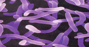
鼠疫耶尔森氏菌(鼠疫菌)是一种需氧不运动、无芽孢、无鞭毛、革兰氏阴性杆菌或球杆菌.革兰氏染色后呈发夹形态,生长温度在4-40°C之间(最适温度28-30°C)。鼠疫菌通过跳蚤在人和啮齿动物间传播,可引起五种主要形式的鼠疫,包括腺鼠疫、败血症鼠疫、肺鼠疫、脑膜鼠疫和咽鼠疫。此外,鼠疫菌可在入侵部位引起皮肤溃疡,表现为痈和溃疡以及脓疱、斑点、瘀点和坏疽。
历史上鼠疫三次大流行已导致全球超过1.6亿人死亡。第一次鼠疫大流行被称为查士丁尼瘟疫,公元541年至750/767年在地中海盆地肆虐,并入侵到北欧、德国和英国。第二次鼠疫大流行从1346年持续到18世纪,包括1346-1353年“黑死病”时期。第三次鼠疫大流行可能始于19世纪末的中国香港,随后通过海上贸易传播到非洲、美洲、大洋洲以及世界其他地区,一直持续到20世纪中叶。目前,鼠疫在世界许多地方仍然存在,自2000年以来被世界卫生组织列为“重新抬头”的传染病。
由于鼠疫菌间同源性较高,血清型和噬菌体分型不适用于鼠疫菌研究。鼠疫菌具有发酵甘油、阿拉伯糖以及还原硝酸盐的能力。基于这些特点可分为五种生物型(antiqua、mediaevalis、orientalis、microtus和Intermedium)。前三种生物型鼠疫菌对动物或人具有高致病性,而田鼠型菌株(包括“pestoides”)对大型哺乳动物无致病性或机会性致病,但对小型啮齿类动物具有毒性。目前,鼠疫菌可划分为五个主要分支33个系统群。其中分支0是鼠疫菌最古老的分支,它包含多个亚群。分支1-4从1330-1340年鼠疫菌“大爆炸”节点中辐射出来。分支1鼠疫菌分布最广,目前在亚洲、非洲和美洲的自然鼠疫疫源地中流行。分支2又分为2.ANT和2.MED,前者在尼泊尔、中国和蒙古传播,后者则遍布亚洲,从高加索、里海、伏尔加-乌拉尔地区,途经哈萨克斯坦西部、土库曼斯坦、吉尔吉斯斯坦北部,一直延伸到中国和蒙古。分支3仅为中国甘肃省、青海省和蒙古菌株,分支4菌株仅在俄罗斯和蒙古被发现。
鼠疫菌含有pCD1质粒(70-75 kb),在进化过程中水平获得了pMT1(100–110 kb)和 pPCP1(9.5 kb)两个质粒,以及鼠疫杆菌独有的由32个基因组成的高致病岛。质粒pMT1和pPCP1编码的一些决定簇有助于鼠疫菌特异性组织侵袭,在跳蚤媒介中存活,以及可能促进其在宿主血液中大量增殖。鼠疫菌中可能的毒力位点(hms、caf1、T3SS和psa)的表达对多种环境胁迫和多种调节蛋白具有响应性。其他负责细胞代谢的基因(包括能量代谢、硫代谢、核糖体蛋白生物合成、铁摄取、血红素合成以及趋化性和运动性)在暴露于多种胁迫时也被激活。黏附侵袭位点是染色体编码的小分子膜蛋白,也是鼠疫菌的主要黏附素。耶尔森菌素对三价铁离子具有高亲和力,是耶尔森氏菌从转铁蛋白和乳铁蛋白中获取铁元素所必需的。
Samples collection date:
Samples host information:
Samples phylogroup information:
Samples Virulence information:
Samples Resistance information:
[1] Perry R D, Fetherston J D. Yersinia pestis - Etiologic agent of plague[J]. Clinical Microbiology Reviews, 1997, 10(1): 35-66.
[2] Lei, Liu, Shangen, et al. Transcriptional regulation of Yersinia pestis biofilm formation[J]. Microbial pathogenesis, 2019, 131: 212-217.
[3] Thompson K M. Environmental Regulation of Yersinia Pathophysiology[J]. Front Cell Infect Microbiol, 2016, 6(25): 25.
[4] Darby C. Uniquely insidious: Yersinia pestis biofilms[J]. Trends in Microbiology, 2008, 16(4): 158-164.
[5] Nuri R, Shprung T, Shai Y. Defensive Remodeling: How bacterial surface properties and biofilm formation promote resistance to antimicrobial peptides[J]. BBA - Biomembranes, 2015: 3089-3100.
[6] Spyrou M A, Bos K I, Herbig A, et al. Ancient pathogen genomics as an emerging tool for infectious disease research[J]. Nat Rev Genet, 2019, 20(6): 323-340.
[7] Barbieri R, Signoli M, Chevé D, et al. Yersinia pestis: the Natural History of Plague[J]. Clin Microbiol Rev, 2020, 34(1): e00044-19.
[8] Qi Z, Cui Y, Zhang Q, et al. Taxonomy of Yersinia pestis[J]. Adv Exp Med Biol, 2016, 918: 35-78.
[9] Zhou D, Han Y, Yang R. Molecular and physiological insights into plague transmission, virulence and etiology[J]. Microbes Infect, 2006, 8(1): 273-84.
[10] Bramanti B, Wu Y, Yang R, et al. Assessing the origins of the European Plagues following the Black Death: A synthesis of genomic, historical, and ecological information[J]. Proc Natl Acad Sci U S A, 2021, 118(36): e2101940118.
[11] Spyrou M A, Tukhbatova R I, Feldman M, et al. Historical Y. pestis Genomes Reveal the European Black Death as the Source of Ancient and Modern Plague Pandemics[J]. Cell Host Microbe, 2016, 19(6): 874-81.
[12] Kutyrev V V, Eroshenko G A, Motin V L, et al. Phylogeny and Classification of Yersinia pestis Through the Lens of Strains From the Plague Foci of Commonwealth of Independent States[J]. Front Microbiol, 2018, 9: 1106.
[13] Han Y, Qiu J, Guo Z, et al. Comparative transcriptomics in Yersinia pestis: a global view of environmental modulation of gene expression[J]. BMC Microbiol, 2007, 7: 96.
[14] Yang R, Atkinson S, Chen Z, et al. Yersinia pestis and Plague: some knowns and unknowns[J]. Zoonoses (Burlingt), 2023, 3(1): 5.