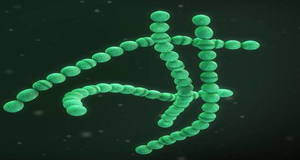
猪链球菌周围被多糖荚膜包围,是一种兼性厌氧、革兰氏阳性、卵状或球状细菌。猪链球菌是一种人畜共患病原菌,可导致猪和人患败血症和脑膜炎。人可以通过密切接触或食用生的、未煮熟的、被污染的猪肉制品而感染猪链球菌。1954年猪链球菌首次被报道,它导致仔猪脑膜炎、败血症、化脓性关节炎的暴发。14年后,丹麦报道了首例人感染猪链球菌的病例。
基于多位点序列分型,猪链球菌可分为1000多种ST型。其中分离自猪的ST1、ST25和ST28菌株最常见,且每种ST型展现出不同的地理优势。猪链球菌种群中存在许多克隆复合体(CCs),引起猪和人感染的最重要的克隆复合体有CC1、CC13/149、CC16、CC17、CC20、CC25、CC28、CC94、CC104、CC233、CC221/234、CC1109、CC1112和CC1237。北美通常为CC25(加拿大)和CC28(美国和加拿大)菌株。CC1菌株在欧洲、亚洲和南美洲有流行;CC20主要分布于荷兰;CC104和CC233(ST233、ST379和ST1656)曾导致猪链球菌在泰国的流行;在欧洲,CC16和CC94中分离自猪的菌株占据主导地位,此外CC94菌株在泰国也引发了人间病例。
血清型鉴定是目前猪链球菌分型的金标准。根据荚膜多糖抗原类型,猪链球菌最初被分为35种血清型(1/2和1–34)。由于其中6种血清型(20、22、26、32、33和34)属于其他细菌,所以官方报道的猪链球菌血清型数量已减少至29种。然而2010年后,中国出现了21/29、NCL21-NCL26和Chz新血清型。其中,血清型2是主要的、可感染人的血清型,能够引发严重的疾病和链球菌中毒性休克综合征(STSS),这也是2005年四川省猪链球菌暴发导致死亡的主要原因。引发STSS的猪链球菌可能通过水平基因转移获得了含有多个毒力基因的89 kbp致病性岛(89 K PAI)。此外,猪链球菌中最常见的毒力标志物还包括muramidase-released protein(MRP)、extracellular protein factor(EF)和hemolysin suilysin(SLY),这些蛋白与血清型2菌株的毒力相关。
目前,已测序的猪链球菌基因组中含有对四环素、大环内酯、氨基糖苷、氯霉素和其他抗菌药物耐药的基因。与其他链球菌相同,猪链球菌中许多耐药基因都是由整合/接合元件、转座子、基因组岛、噬菌体和嵌合元件携带的。据报道,猪链球菌菌株对青霉素和氨苄西林敏感;高达85%的菌株对大环内酯类、林克酰胺类、四环素类和磺胺类药物具有耐受性。自2010年以来,人感染猪链球菌的病例数大幅提升,且大多病例来自于东南亚地区。
Samples collection date:
Samples host information:
Serotype information:
Samples MLST information:
Samples Virulence information:
Samples Resistance information:
[1] Brizuela J, Kajeekul R, Roodsant TJ, et al. Streptococcus suis outbreak caused by an emerging zoonotic strain with acquired multi-drug resistance in Thailand. Microb Genom. 2023;9(2):mgen000952.
[2] Wertheim HF, Nghia HD, Taylor W, Schultsz C. Streptococcus suis: an emerging human pathogen. Clin Infect Dis. 2009;48(5):617-625.
[3] Nicholson TL, Kalalah AA, Eppinger M. Population structure and genetic diversity of Streptococcus suis isolates obtained from the United States. Front Microbiol. 2023;14:1250265.
[4] Hatrongjit R, Fittipaldi N, Gottschalk M, Kerdsin A. Tools for Molecular Epidemiology of Streptococcus suis. Pathogens. 2020;9(2):81.
[5] Scherrer S, Rosato G, Spoerry Serrano N, et al. Population structure, genetic diversity and pathotypes of Streptococcus suis isolated during the last 13 years from diseased pigs in Switzerland. Vet Res. 2020;51(1):85.
[6] Fittipaldi N, Xu J, Lacouture S, et al. Lineage and virulence of Streptococcus suis serotype 2 isolates from North America. Emerg Infect Dis. 2011;17(12):2239-2244.
[7] Segura M, Aragon V, Brockmeier SL, et al. Update on Streptococcus suis Research and Prevention in the Era of Antimicrobial Restriction: 4th International Workshop on S. suis. Pathogens. 2020;9(5):374.
[8] Athey TB, Teatero S, Takamatsu D, et al. Population Structure and Antimicrobial Resistance Profiles of Streptococcus suis Serotype 2 Sequence Type 25 Strains. PLoS One. 2016;11(3):e0150908.
[9] Schultsz C, Jansen E, Keijzers W, et al. Differences in the population structure of invasive Streptococcus suis strains isolated from pigs and from humans in The Netherlands. PLoS One. 2012;7(5):e33854.
[10] Brizuela J, Kajeekul R, Roodsant TJ, et al. Streptococcus suis outbreak caused by an emerging zoonotic strain with acquired multi-drug resistance in Thailand. Microb Genom. 2023;9(2):mgen000952.
[11] Kerdsin A, Hatrongjit R, Gottschalk M, et al. Emergence of Streptococcus suis serotype 9 infection in humans. J Microbiol Immunol Infect. 2017;50(4):545-546.
[12] Obradovic MR, Segura M, Segalés J, Gottschalk M. Review of the speculative role of co-infections in Streptococcus suis-associated diseases in pigs. Vet Res. 2021;52(1):49.
[13] Wang X, Sun J, Bian C, et al. The population structure, antimicrobial resistance, and pathogenicity of Streptococcus suis cps31. Vet Microbiol. 2021;259:109149.
[14] Tan C, Zhang A, Chen H, Zhou R. Recent Proceedings on Prevalence and Pathogenesis of Streptococcus suis. Curr Issues Mol Biol. 2019;32:473-520.
[15] Varela NP, Gadbois P, Thibault C, Gottschalk M, Dick P, Wilson J. Antimicrobial resistance and prudent drug use for Streptococcus suis. Anim Health Res Rev. 2013;14(1):68-77.
[16] Susilawathi NM, Tarini NMA, Fatmawati NND, et al. Streptococcus suis-Associated Meningitis, Bali, Indonesia, 2014-2017. Emerg Infect Dis. 2019;25(12):2235-2242.